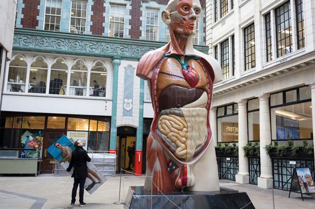
It is a rite of passage for students dating back centuries, but some medical schools are turning away from cadaveric dissection as costs spiral – although many believe technological alternatives still cannot compete with the experience of coming face to face with a corpse.
The University of Sheffield has told staff that it has been unable to resolve “ventilation issues” that have prevented its medical school using “wet specimens” for much of the past academic year and it will now “transition away from teaching human anatomy through cadaveric dissection on a permanent basis”, according to an email from head of faculty Ashley Blom, seen by Times Higher Education.
Professor Blom said teaching will now take place using “a combination of prosections, plastinates and models” while the university “takes inspiration from leading anatomy programmes around the world” to devise longer-term plans for its human anatomy teaching.
Campus resource: Generation Z presents new challenges for medical education
He cited the fact that many other universities have also adopted alternative methods to cadaveric dissection and asserted that the move “won’t alter the overall learning outcomes of our programmes”.
The University of Oxford has also stopped using cadaveric dissection in its medical school, while it is also not offered by universities including Exeter and Plymouth; at others, such as Hull York Medical School, it is used only on certain courses.
Research published in 2021 found that 34 of 39 UK medical schools polled still offered full body dissection in 2019, but it is thought that the Covid pandemic exacerbated moves towards virtual alternatives.
One of this study’s authors, Claire Smith, professor of anatomy at Brighton and Sussex Medical School, said the decision was often motivated by health and safety reasons, or saving estate costs.
“Running a donor facility is an expensive operation,” she said. “And it’s difficult to do, given the ageing architecture of many universities.”
Virtual reality, videos and ultrasound machines are increasingly being used in anatomy teaching, but Professor Smith said she still saw these as adjuncts to cadaveric dissection.
“It is part of the mix, but none of these simulations are there yet to completely replicate the dissection experience,” said Professor Smith, who was featured in the recent Channel 4 television documentary My Dead Body, which showed the dissection of a young woman who had died from cancer.
“If you are trying to learn purely anatomical content, you can do that from virtual and digital sources. What is hard to learn is the spatial relation – understanding if an artery feels squidgy or soft, what a nerve feels like. You can qualify as a doctor without ever having practised through dissection, but I think it adds a real value to that learning,” she said.
“The donor is a student’s first patient, so it also adds a lot of humanity to the teaching. This is a person who laughed and cried, had family, friends.”
Professor Smith added that it was notable that some of the new medical schools that have opened in recent years – Anglia Ruskin and Kent and Medway – had chosen to invest in donor facilities but, at the schools that have moved away from it, cadaveric dissection “will be incredibly hard to reinstate”.
“When you think about training as a doctor, there are some very strong rites of passage that you know you are going to be learning, and dissection is seen as part of a tradition in medicine,” she said. “But it also has a very real application to effective and safe patient care today.”
Others in the field stressed that simulations can provide a teaching experience that is possibly more effective than using cadavers.
“Different educational formats and methods to teach anatomy will be appropriate at the various stages of medical training,” said Frank Smith, professor of vascular surgery and surgical education at the University of Bristol, and a council member at the Royal College of Surgeons of England.
He added that physical simulations and digital platforms were now “very effective” and “improving all the time”, so can “provide a more targeted teaching experience than cadavers can”.
“Where available and permitted, the use of cadaveric materials for undergraduate teaching is a useful tool, particularly for those students who are interested in surgery,” Professor Smith said.
“There needs to be equity of access in undergraduate medical education. Some students will not get exposure to cadaveric materials during their studies, so it is important to acknowledge the range of ways this can be done successfully now.”
Register to continue
Why register?
- Registration is free and only takes a moment
- Once registered, you can read 3 articles a month
- Sign up for our newsletter
Subscribe
Or subscribe for unlimited access to:
- Unlimited access to news, views, insights & reviews
- Digital editions
- Digital access to THE’s university and college rankings analysis
Already registered or a current subscriber? Login


